


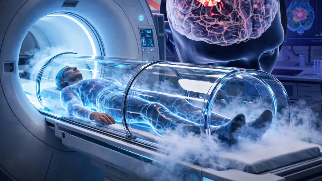
One of the most compelling advances in modern oncology is emerging from Liverpool Hospital in south-west Sydney, Australia, where clinicians have introduced the country’s first fully MRI-guided cryoablation system. This cutting-edge, minimally invasive cancer treatment is redefining how doctors target select tumors, using extreme cold to destroy cancerous tissue with millimeter-level precision while the patient remains inside an MRI scanner. No scalpels. No stitches. Instead, MRI-guided cryoablation freezes tumors from the inside out, turning imaging technology into a therapeutic weapon.
Below, we break down the science behind this approach, walk through the procedure step by step, examine its clinical applications, benefits, and limitations, and explore what it signals for the future of precision cancer care.
Understanding Cryoablation: The Basics of Freezing Tumors
Cryoablation, at its core, is a technique that uses extreme cold to kill abnormal cells. The term “cryo” comes from the Greek word for cold, and “ablation” means removal or destruction. This method has been around for decades, initially used in dermatology to freeze off warts or in cardiology to treat irregular heart rhythms. In oncology, it’s applied to destroy tumors by disrupting cellular structures freezing causes ice crystals to form inside cells, rupturing membranes and cutting off blood supply, leading to cell death.
What makes the Sydney implementation stand out is the integration with magnetic resonance imaging (MRI). Traditional cryoablation often relies on ultrasound or CT scans for guidance, but MRI provides superior soft-tissue contrast and real-time imaging without ionizing radiation. This allows doctors to visualize the tumor and surrounding tissues in exquisite detail, ensuring the freeze zone targets only the malignancy while sparing healthy structures like nerves, blood vessels, or organs.
The freezing agent is typically argon gas or liquid nitrogen, delivered through a thin needle called a cryoprobe. When the gas expands inside the probe, it creates temperatures as low as -160°C (-256°F), forming an “iceball” a spherical zone of frozen tissue that expands to encompass the tumor. The process involves cycles of freezing and thawing: freezing kills the cells, while thawing helps confirm the destruction and allows for multiple probes if needed for larger tumors.
The Role of MRI In Real-Time Precision in Action
MRI-guided cryoablation elevates this to a new level of sophistication. MRI machines use powerful magnets and radio waves to generate detailed 3D images of the body’s interior, excelling at distinguishing between soft tissues like muscles, fat, and tumors something CT scans struggle with due to similar densities.
During the procedure, clinicians position the patient inside a specialized MRI scanner designed for interventional use. The scanner has an open configuration or built-in modifications. These allow probe insertion without disrupting the magnetic field.
Real-time MRI sequences provide continuous feedback, updating every few seconds to show the probe’s path and the growing iceball. This is crucial because the iceball appears as a dark void on MRI images, making it easy to monitor its size and shape typically aiming for a margin of 5-10 mm beyond the tumor to ensure complete coverage.
At Liverpool Hospital, this system is described as one of the most advanced on Earth for this purpose. It’s particularly effective for tumors in tricky locations, such as the spine, pelvis, or soft tissues, where surgery could risk paralysis or severe complications. The MRI’s precision minimizes damage to adjacent structures, reducing side effects like nerve pain or organ dysfunction.
Step-by-Step: How the Procedure Unfolds
Let’s walk through how clinicians perform the procedure at facilities like Liverpool Hospital, drawing on detailed procedural descriptions.
- Preparation and Diagnosis: Before the procedure, patients undergo comprehensive imaging. This often includes a combination of CT, MRI, or ultrasound. These images map the tumor’s size, location, and characteristics. Biopsies may confirm it’s cancerous.
- Eligibility is key: This treatment suits small to medium tumors (typically under 5 cm). It is suitable for patients unfit for surgery due to age, comorbidities, or tumor location. It also helps those seeking pain relief from metastatic cancer. Anesthesia options include conscious sedation or general anesthesia, depending on the site.
- Probe Insertion: The patient is in the MRI scanner. Interventional radiologists make a tiny skin puncture under local anesthesia. The puncture is about 2-3 mm. Using real-time MRI guidance, they advance the cryoprobe—a slender needle about 1.5-2.5 mm in diameter directly into the tumor. For larger tumors, multiple probes (up to 4-6) are placed strategically to overlap iceballs.
- Freezing Cycles: Once positioned, argon gas flows through the probe, rapidly cooling the tip. The first freeze lasts 8-10 minutes, forming the iceball. MRI monitors its expansion in real time, ensuring it engulfs the tumor without encroaching on vital areas. A thaw phase (3-5 minutes) follows, using helium gas to warm the probe, allowing ice to melt and confirming cell death. This cycle repeats 1-2 times for thorough destruction.
- Completion and Recovery: Probes are removed, and the puncture site is bandaged—no stitches needed. The entire session takes 1-3 hours. Patients often go home the same or next day, with minimal downtime compared to surgery, which might require weeks of recovery.
Over the following weeks, the body absorbs the dead tissue, and follow-up scans confirm tumor shrinkage.
Game-Changer for Pain Management and Precision Oncology
The advantages are compelling. First, it’s outpatient-friendly: no hospital stay, lower infection risk, and quicker return to normal life. For pain relief, it’s transformative—patients with bone or soft-tissue metastases often experience immediate reduction in debilitating pain, as the freezing destroys nerve endings in the tumor.
Precision is another hallmark. MRI’s millimeter-level accuracy (down to 1 mm) allows treatment of tumors near critical structures, like the spinal cord, where surgery is risky. Studies show success rates of 85-95% for complete tumor ablation in suitable cases, with lower recurrence compared to some alternatives.
It’s especially valuable for kidney, liver, prostate, breast, and bone tumors, and for palliative care in advanced cancers. In Sydney, clinicians and patients alike hail the approach as a breakthrough, citing cases such as a 64-year-old grandmother who woke pain-free after years of sleepless nights caused by spinal tumors.
Risks and Limitations
No treatment is without downsides. Potential complications include bleeding, infection, or damage to nearby tissues if the iceball expands too far though MRI minimizes this. Some patients experience temporary pain, swelling, or “cryoshock” (a rare systemic reaction from large tumor destruction). It’s not ideal for very large tumors (>5 cm) or those in highly vascular areas, where bleeding could be an issue.
Long-term data is still emerging; while effective for local control, it may not address metastatic spread, often requiring combination with chemotherapy or radiation. High costs and limited availability remain major barriers, as specialized MRI suites drive expenses and confine access to major centers like Liverpool Hospital.
Real-World Impact: Stories from Sydney
At Liverpool Hospital, this technology has already changed lives. One reported case involved treating spinal tumors causing severe, chronic pain. Post-procedure, patients report dramatic relief, avoiding opioids or invasive surgeries. Clinicians note it’s particularly suited for Australia’s aging population, where surgical risks are higher.
Globally, institutions like Stanford Health Care have similar programs, reporting fewer complications than surgery and high patient satisfaction.
The Future of Non-Invasive Cancer Warfare
This Sydney launch signals a shift toward “interventional oncology,” where imaging and targeted therapies converge. Ongoing research explores combining cryoablation with immunotherapy—freezing tumors may release antigens that boost the immune response against cancer elsewhere in the body. Advances in probe design and AI-assisted MRI could make it even more precise and accessible.
In essence, MRI-guided cryoablation isn’t just treating cancer; it’s redefining it as a battle won with cold calculation rather than cuts. As more hospitals adopt this, it could become a standard for early-stage tumors, offering hope to millions. If recent journals are any indication, we’re on the cusp of a colder, kinder era in oncology.
Reference
Kohli A, Hui T, Osman S, et al. Image-guided percutaneous cryoablation for hepatic tumours: Efficacy and safety in a retrospective cohort. J Med Imaging Radiat Oncol. 2025;69(5): doi:10.1111/1754-9485.70008.