Cells or organs of the body which detect stimuli are called receptors or sense organs. Every individual cell of protozoans and in sponges is sensitive to such changes like light intensity, concentration of chemicals, temperature changes and water current etc.
RECEPTORS (SENSE ORGANS)
Ectodermal sensory cells or receptor cells develop in cnidarians for the first time and they regularly occur in all metazoans. Sense organs have dual functions (i) they detect environmental changes or stimuli and (ii) transmit this information to the central nervous system.
Types of Receptors According to Location
Receptors may be classified according to the location in the body:
1. Exterocepters
The receptors situated on the surface of body are called exteroceptors. These receive environmental stimuli from outside like touch, pressure, taste, heat, etc. These include eyes, ears, nose, taste buds and cutaneous sense organs.
2. Proprioceptors
These are stretch receptors present in the muscles, joints, tendons, connective and skeletal tissues. They supply information about the kinesthetic sense of equilibrium and orientation. They are responsible for maintenance of body posture.
3. Interoceptors
The receptors present in internal organs are called interoceptors. They provide information about the internal changes in body environment, such as CO₂ concentration. blood composition, painfulness, etc. The stimulus of hunger, thirst, urination, suffocation etc., originates in internal organs of the body.
EYE (PHOTORECEPTOR)
[1] External features
1. Shape and size
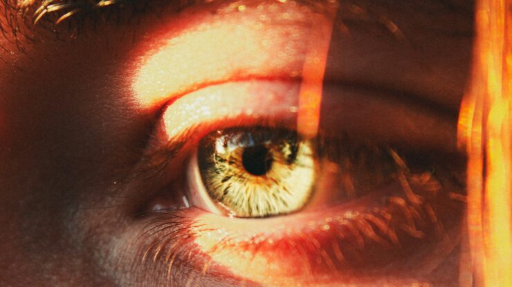
The human eyeball is hollow, spherical organ measuring about 2.5 cm in diameter. The anterior one fifth is exposed while four fifth part remains concealed within eye orbit.
2. Eye lids
Movable upper and lower eye lids can close the eye by orbicularis oculi muscle, when required and protect it from dust particles or other objects. The stiff hair or eyelashes present on the free edges of eyelids also guard the eye against dust particles, rain, sweat and glare. The nictitating membrane is vestigial in human eye and represented by a pink semilunar mass, called plica semilunaris.
3. Eye brows
The richly hairy skin surrounding the human eyes occur as two thick and arched eminences called eye brows. They provide further protection to the eye and are developed in primates, mainly in human.
4. Extraocular muscles
Movements of the eye ball in different directions within its orbit are brought about by six voluntary strap shaped ocular or eye ball muscles. The ocular muscles are superior, inferior, external and internal rectus and superior and inferior oblique. The superior oblique muscle moves eye ball downward and outward, while inferior oblique moves it upward and outward. The superior rectus moves eyeball upward and inward whereas inferior rectus moves it downward and inward .
5. Glands
The oily secretions of Melbomian and Harderian glands lubricate the eyelids and cornea. The lacrimal gland is present under the upper eyelid towards its outer corner. It secretes a slightly saline and antiseptic watery fluid, the tears. Harderian gland is present in some mammals like shrews, whales and mice but not in man. Glands of Moll are also found in human eye which open into the follicles of eye lashes to lubricate them.
[2] Internal structure
A cross section reveals that the eyeball is hollow from within. Its wall is made of three coats or layers of tissue in close contact: an outermost sclerotic, middle choroid and the innermost retina .
1. Sclerotic
The sclerotic coat is made of dense connective tissue, collagen fibres giving strength to the wall. Its small anterior exposed part is transparent, through which light enters, and is called cornea. It bulges out slightly and its external surface is intimately covered with a thin, transparent epidermal layer, the conjunctiva. The conjunctiva is the thinnest layer in eye and it represents the thinnest part of the skin.
2. Choroid
The middle layer choroid is a thin connective tissue layer heavily pigmented and richly vascular. The choroid lines only the muscles. posterior region of sclera. Anteriorly, the choroid enlarges to from the ciliary body. It contains ciliary muscles and ciliary processes, which secrete the aqueous humor.
In front of the ciliary body, the choroid forms the iris. It is visible from outside as an opaque brown disc like diaphragm perforated in the centre by a round hole, the pupil. The iris contains smooth muscles. The different colours of eye in different persons like black, brown, grey, blue or green is due to heavy pigmentation in iris. The iris of eye can be compared with the diaphragm of a camera as it regulates the amount of light entering in eye by constricting or dilating the pupil.
3. Retina.
It is the innermost, neuro- sensory layer of the eyeball sensitive to light.
(a) Histology of retina
The retina consists of an outer non-nervous pigmented layer closely applied with the choroid, and the inner nervous layer containing two main types of sensory cells, rods and cones, named because of the characteristic shapes of their receptor terminations. When stimulated by light, they generate electric impulses and relay the information to the brain by way for the optic nerve.
In a V.S. of retina, the various layers forming it from outside are
(i) pigment epithelium,
(ii) receptive layer of rods and cones,
(iii) bipolar neurons,
(iv) ganglion cells and
(v) layer of nerve fibres which converge to form optic nerve
(a) Rods
They are more sensitive to light of low intensity. They are suitable for night vision but the image formed is diffused, lacking in details.
(b) Cones
They are sensitive to light of high intensity. They are more suited for day vision and they produce a sharp image with fine details. They are associated with appreciation of colours. Colour perception in mammals is confined to man and other primates only. Human eye has about 115 million rods and about 6.5 million cones.
Cones are more concentrated towards the centre while rods are more numerous towards the periphery. The spot where all the nerve fibres converge to form optic nerve and it leaves the eye is called the blind spot no image is formed here. Near it lies the yellow spot or macula lutea containing only cones filled with yellow pigment. This part has fovea centralis marked by a slight oval depression along the optical or visual axis. It is the point of principal focus or brightest image.
Inverted retina
The rods and cones setup – impulses of photoreception which pass in reverse direction through bipolar neurons to reach optic nerve. This arrangement is referred to as inverted retina.
4. Lens
A crystalline, solid and biconvex lens is present just behind the pupil. It is held in position by suspensory ligaments or zonula of Zinn which are radially arranged fibres connecting the lens with the ciliary processes. In old age some people suffer from cataract, in which the lens gradually becomes opaque causing blindness. It is cured by replacing the lens.
5. Chambers
The iris, lens and its suspensory ligaments divide the internal cavity of eye into two unequal compartments. Between the lens and the cornea is the smaller anterior aqueous chamber. It is filled with a clear lymph-like watery fluid, the aqueous humor containing about 98% water, protein, glucose and NaCl. It is secreted continually by the ciliary body and drained through canal of Schlemm into the blood at the base of the cornea. Aqueous humor nourishes cornea and lens and maintains intraocular pressure.
Between the lens and the retina is the larger posterior vitreous chamber, filled with a gelatinous secretion, the vitreous humor. This substance mainly contributes to intraocular pressure and prevents eye ball from collapsing.Vitreous humor unlike the aqueous humor, does not undergo consistent replacement. It is essentially protein-gel having high viscosity due to presence of hyaluronic acid. An imbalance in the intraocular pressure leads to a disease called glaucoma in which retina is injured.
Working of Eye
1. Image formation
There is a close similarity in the structure and working of an eye to a photographic camera (Fig. 16). A camera can be opened or shut by its shutter, the eye by its lids. Both have convex lenses (cornea + lens in eye) which focus light rays on a sensitive film (retina of eye) forming an inverted image. In both, the amount of light entering the chamber is controlled by diaphragm (iris). The interior of a camera is painted black to avoid internal reflections which might blur the image. The eye also has a highly pigmented lining, the choroid coat. Finally the camera can be focussed by moving the lens to and fro, and the eye can also accommodate to see near and far objects.
2. Photochemistry of vision
The rods of mammalian eye contain an extremely photosensitive substance called rhodopsin or visual purple. It is a compound of opsin or scotopsin (protein) and retinal or retinene (pigment). During bright light it disappears. It breaks up into opsin and retinene in dim light. In darkness opsin and retinene recombine to form rhodopsin with the aid of ATP. Retinene is a derivative of vitamin A.
On coming out of a dark cinema hall in bright day light, one feels dazzled until the level of rhodopsin is reduced in the rods. An acute deficiency of vitamin A in the body may result in night blindness, that is, difficulty in seeing in dim light.
3. Focusing or accommodation
The ability to sharply focus or clearly see objects placed at various distances is known as accommodation. The eye does so by changing the shape or curvature of its elastic lens (Fig. 17 and 18).
4. Colour vision
The light sensitive pigment of cones is iodopsin or visual violet. There are three kinds of cones which are stimulated by different wavelengths of light corresponding to the blue, green and red parts of the spectrum. This hypothesis is supported by the three known types of colour blindness.
The human eye can recognize about 150 different colours in the visible spectrum and can detect differences of less than 3 mµ in wavelength.
The erythrolabe, chlorolabe and cyanolabe are three different types of photopigments in cones which make these cones selectively sensitive to the red, green and blue colours, respectively .
A person with loss of red cones is known as a protonope, the colour blind person who lacks green cones is called a deuteranope while a man with an insensitivity to blue colour is known as tritanope.
5. Binocular (stereoscopic) vision
In man, other primates and birds of prey like owls and eagles etc., both the eyes face forward, so that they form two separate images of the same object. But two images are not seen due to overlapping of their fields of vision. As a result, the object is seen in three dimensions which also gives a sense of distance of the object in view. This type of vision in which the distance is also judged is called binocular or stereoscopic vision. Minimum distance for binocular vision in human eye is 25 cm. The human eye is sensitive to light rays of wave lengths from 380 to 760 nanometers or about 4000Å to 7000Å and it is most sensitive to light rays of 5000Å.
6. Tapetum layer
In many animals the inner layer of choroid next to retina, is modified to reflect light rays falling upon it through retina and the eye appears to shine or glare in the dark.

