"CONNECTING RESEARCH TO REALITY"

Oxidative Phosphorylation

Explore the intricate process of oxidative phosphorylation , essential for cellular energy production. Delve into its mechanisms, biological significance, and implications with our detailed and informative guide.

OXIDATIVE PHOSPHORYLATION

Shibasis Rath

7/7/20249 min read

Principles of Bioenergetics-Oxidative Phosphorylation

1.Conservation of Energy (First Law of Thermodynamics):

• Energy cannot be created or destroyed in any physical or chemical change.

• It can only be transferred from one form to another.

Example:

In a biological system, the energy in food (chemical energy) is converted into mechanical energy and heat by cells.

2.Second Law of Thermodynamics:

• States that in any energy transformation, there is an increase in the disorder (entropy) of the universe.

• Implication for Living Organisms:

• Living cells constantly exchange matter and energy with their surroundings.

• They are never in complete equilibrium, as they are perpetually growing, dividing, and maintaining internal order (which increases entropy in their surroundings).

3.Gibbs Free Energy (ΔG):

• ΔG determines whether a chemical reaction will occur spontaneously.

• ΔG < 0: Reaction is exergonic (spontaneous and releases energy).

• ΔG > 0: Reaction is endergonic (requires energy input to proceed).

• Significance: Cells use ΔG to drive metabolic processes essential for life.

4.Enthalpy (ΔH):

• The total heat content of a system undergoing a chemical reaction.

• Exothermic reactions release heat (ΔH < 0); endothermic reactions absorb heat (ΔH > 0).

• Role in Bioenergetics: Enthalpy changes accompany biochemical reactions such as digestion and cellular respiration.

5.Entropy (ΔS):

• Measures the randomness or disorder in a system.

• ΔS > 0: Increase in disorder (spontaneous processes tend towards higher entropy).

Mitochondrial Electron Transport

• Mitochondria are double-membraned organelles with an outer membrane and an inner membrane, which encloses the matrix.

• The inner membrane is highly folded into structures called cristae, increasing its surface area.

• These folds contain protein complexes crucial for electron transport and oxidative phosphorylation.

☀ Outer Membrane:

• Porous and permeable, allows passage of small molecules and ions.

☀Inner Membrane:

• Impermeable to most ions and small molecules; regulates passage via specific transport proteins.

• Contains electron transport chain complexes and ATP synthase, essential for oxidative phosphorylation.

☀Cristae:

• Folds of the inner membrane that house electron transport chain components.

• Increase surface area for ATP production through oxidative phosphorylation.

☀Electron Transport Chain (ETC):

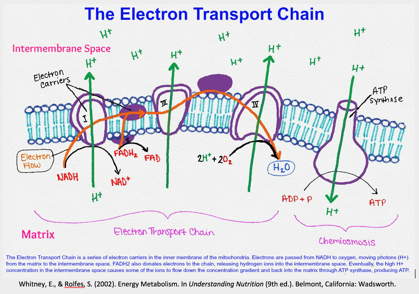
• Series of protein complexes (I, II, III, IV) embedded in the inner membrane.

• Facilitates electron transfer from NADH and FADH2 to oxygen, generating ATP.

☀Oxidative Phosphorylation:

• Process where ATP is synthesized using energy released by electron transport.

• ATP synthase uses the proton gradient across the inner membrane (created during electron transport) to drive ATP production.

• Produces the majority of ATP in aerobic cells through oxidative metabolism.

• Essential for cellular energy production and maintenance of metabolic homeostasis.

• Mitochondrial electron transport and oxidative phosphorylation are vital for cellular respiration, providing energy for various biological processes and maintaining cellular function.

Mitochondrial Electron Transport Chain and Cellular Respiration:

• The mitochondrial electron transport chain, also known as the respiratory chain, consists of four main complexes:

1.Complex I (NADH-Coenzyme Q Reductase):

Catalyzes the transfer of electrons from NADH to Coenzyme Q (Ubiquinone).

2.Complex II (Succinate-Coenzyme Q Reductase):

Catalyzes the transfer of electrons from FADH₂ to Coenzyme Q.

3.Complex III (Cytochrome bc₁ Complex):

Transfers electrons from Coenzyme Q to Cytochrome c.

4.Complex IV (Cytochrome c Oxidase):

Catalyzes the final transfer of electrons to molecular oxygen (O₂), producing water (H₂O).

• During cellular respiration, which includes glycolysis, the citric acid cycle (Krebs cycle), and oxidative phosphorylation, glucose is completely oxidized to CO₂ and H₂O.

• The electron carriers NADH and FADH₂ generated during glycolysis and the citric acid cycle donate electrons to the electron transport chain (ETC) embedded in the inner mitochondrial membrane.

• As electrons move through the ETC, they pump protons (H⁺ ions) from the mitochondrial matrix to the intermembrane space, establishing a proton gradient.

• This gradient drives ATP synthesis through ATP synthase, a process known as oxidative phosphorylation.

• ATP is the primary energy currency of the cell and is essential for cellular functions and processes.

• Understanding the mitochondrial electron transport chain and cellular respiration is crucial for comprehending how cells generate energy from nutrients like glucose under aerobic conditions.

Respiratory Complex- I (NADH-CoQ Reductase Complex)

• Complex I is the first and largest enzyme complex in the mitochondrial electron transport chain. It plays a crucial role in catalyzing the transfer of electrons from NADH to coenzyme Q (CoQ), also known as ubiquinone. Here’s a structured overview:

☀ Structure and Components:

• Contains multiple subunits including flavin mononucleotide (FMN) and iron-sulfur clusters (Fe-S).

• Consists of several iron-sulfur proteins that facilitate electron transfer.

☀ Function:

• Catalyzes the transfer of electrons from NADH to CoQ, generating CoQH₂ (reduced form of CoQ) and NAD⁺ (oxidized form of NADH).

• This process is critical for the generation of a proton gradient across the mitochondrial inner membrane, essential for ATP synthesis.

☀ Electron Transfer reaction:

Steps :

1. NADH binds to Complex I, donating electrons.

2. Electrons pass through FMN (which becomes FMNH₂, then oxidized back to FMN).

3. Electrons are transferred through a series of iron-sulfur clusters within the complex.

4. Ultimately, electrons are transferred to CoQ, reducing it to CoQH₂.

5. This transfer of electrons is coupled with the translocation of protons across the inner mitochondrial membrane, contributing to the proton gradient.

☀ Role in ATP Production:

• Complex I initiates the flow of electrons down the electron transport chain, leading to ATP synthesis by ATP synthase.

• ATP synthesis occurs as protons flow back into the mitochondrial matrix through ATP synthase, driven by the proton gradient established by Complex I and other complexes.

• Complex I is essential for cellular respiration, playing a fundamental role in energy production through oxidative phosphorylation. Understanding its structure and function is crucial for comprehending mitochondrial function and energy metabolism.

☀ Electron Transport Chain and Acceptors:-

• Pro NADH:Participates in two-electron transfer.

• FMN and CoA:Can accept and donate electrons, facilitating the transfer of these electrons.

• Receive electrons from reduced coenzymes and undergo one-electron reduction.

• Coenzyme Q (CoQ):

- Acts as a mobile electron carrier.

- Its hydrophobic nature allows it to reside and move easily within the lipid bilayer of the mitochondrial membrane.

Respiratory Complex II

• Complex II, also known as succinate dehydrogenase (SDH), is an integral membrane protein complex located in the inner mitochondrial membrane.

• It plays a crucial role in both the electron transport chain and the citric acid cycle (Krebs cycle).

Function:

• Complex II catalyzes the oxidation of succinate to fumarate in the citric acid cycle and simultaneously transfers electrons to the electron transport chain.

☀ Components:

✓Succinate Dehydrogenase (SDH): The enzymatic core of Complex II, containing the FAD (Flavin Adenine Dinucleotide) cofactor.

✓Iron-Sulfur (Fe-S) clusters: These clusters facilitate electron transfer from succinate to ubiquinone (coenzyme Q, CoQ).

✓Cytochrome b: Part of the electron transport chain within Complex II, facilitating electron transfer.

☀ Electron Transfer:

1. Succinate is oxidized to fumarate by succinate dehydrogenase, releasing electrons.A covalent bonds found.

2. Electrons are transferred to iron-sulfur clusters within Complex II.

3. Finally, electrons are passed to coenzyme Q (CoQ), which continues their transport through the electron transport chain.

☀ Chemical Reaction:

• The chemical reaction catalyzed by succinate dehydrogenase is:

Succinate + FAD→Fumarate + FADH^2

• This reaction releases electrons that are transferred through Complex II to CoQ, contributing to the generation of ATP through oxidative phosphorylation.

☀ Proton Pumping:

• No proton pumping across the membrane is associated with complex -ll.

• This means no protons are pumped during the electron transfer reaction in this complex.

Complex III (Cytochrome bc1 Complex)

✓ CoQH^2 + 2cy + c [Fe (III)]→ CoQ+2cy +Fe(ll)+2H+

• Complex III, also referred to as the Cytochrome bc1 complex, plays a crucial role in the electron transport chain of mitochondria.

☀ Location and Function:

• Located within the inner mitochondrial membrane.

• Facilitates electron transfer during oxidative phosphorylation.

☀ Components:

- Two b-type cytochromes.

- One cytochrome c1.

- One Fe-3 cluster.

☀ Cytochromes:

• Contains redox-active proteins including cytochromes.

• Cytochromes alternate between Fe(4) and Fe(1) oxidation states during electron transport.

• Redox-active proteins found in almost all organisms.Contain heme groups that alternate between Fe (4) and Fe (3) oxidation states during electron transport.

• Reduced cytochrome C (Fe (2+) state) exhibits characteristic absorption spectra with distinct peaks known as Bovet bands.

• The wavelength of the d-peak varies among different cytochromes.

• The d-peak is absent in oxidized cytochromes.

• Common types include cytochrome a, cytochrome b, and cytochrome c.

• Variations exist within each type leading to different subtypes.

☀ Electron Transfer Process:

• Electrons received from ubiquinol (CoQH2) and cyt c [Fe(III)].

• Transfers electrons to cytochrome c (cyt c) and generates a proton gradient (H+) across the inner mitochondrial membrane.

• Reduced cytochrome c (Fe(1) state) exhibits distinct absorption spectra with characteristic peaks (Bovet bands).

• Variations in peak wavelengths (e.g., d-peak) among different cytochromes reflect subtype differences.

☀ Asymmetric Location:

• Complex III is asymmetrically located within the inner mitochondrial membrane.

• Cytochrome c and the non-heme iron-sulfur (Rieske) protein are positioned towards the outer surface.

• Cytochrome b spans through the membrane.

☀ Role of Cytochrome C:

• Cytochrome c is a small protein loosely bound to the outer surface of the inner mitochondrial membrane.

☀ Functions:

It functions to shuttle electrons:

• From the heme c of Complex III to cytochrome c oxidase (Complex IV) by transiently binding there.

• Cytochrome c accepts electrons from Complex III, facilitating the reduction of oxygen to water at Complex IV.

• The transfer of electrons from Complex II to Complex III requires only one electron, whereas the reduction of oxygen to water at Complex IV requires two electrons.

• Thus, for every molecule of coenzyme Q, two molecules of cytochrome c are needed.

Respiratory complex lV :-

4 Cytc [Fe(II)] + 4 cyt c [Fe(II)] + O₂ → 4 Cytc [Fe(III)] + 4 cyt c [Fe(III)] + 2H₂O

Cytochrome c oxidase, also known as Complex IV, is a crucial enzyme in the mitochondrial electron transport chain, facilitating the final step where molecular oxygen is reduced to water. Here are detailed aspects of its structure, function, and mechanism:

1. Structure and Composition:

- Cytochrome c oxidase is located in the inner mitochondrial membrane and consists of multiple subunits. Key components include:

- Heme groups: Specifically, cytochromes a and a₃, which are involved in electron transfer.

- Copper ions: Two copper centers, CuA and CuB, which also play critical roles in electron transfer and oxygen reduction.

2. Function:

- Electron Transport: Cytochrome c oxidase catalyzes the transfer of electrons from reduced cytochrome c molecules to oxygen (O₂). It sequentially oxidizes four molecules of reduced cytochrome c (Cytc [Fe(II)]) while simultaneously reducing one molecule of O₂.

- Oxygen Reduction: The reduction of molecular oxygen involves a four-electron reduction process, ultimately producing two molecules of water (2H₂O).

- Proton Pumping: During the electron transfer process, cytochrome c oxidase facilitates the pumping of protons (H⁺) across the inner mitochondrial membrane. This proton pumping generates an electrochemical gradient, which is essential for ATP synthesis in oxidative phosphorylation.

3. Mechanism:

- Active Site: The active site of cytochrome c oxidase, located near the cytochrome a₃ and CuB centers, facilitates the binding and reduction of oxygen. It involves a series of redox reactions where electrons are transferred from Cytc [Fe(II)] to the heme groups and copper centers, ultimately leading to the reduction of O₂ to water.

- Proton Pumping Mechanism: Protons are pumped across the inner mitochondrial membrane during the reduction of oxygen. This process is coupled to electron transfer and involves conformational changes in the enzyme complex.

4. Biological Significance:

- Cytochrome c oxidase is essential for cellular respiration, as it is the terminal enzyme of the electron transport chain. It ensures efficient energy production by converting the energy stored in reduced cytochrome c and molecular oxygen into a proton gradient, which drives ATP synthesis.

- Mutations or deficiencies in cytochrome c oxidase can lead to mitochondrial diseases and impaired cellular respiration, affecting various tissues and organs.

Cytochrome c oxidase plays a central role in mitochondrial function and energy metabolism. Its complex structure and precise mechanism ensure the efficient reduction of oxygen to water, coupled with the generation of a proton gradient critical for ATP production in cells.

Inhibitors of electron transport chain (ETC)

• These are substances that interfere with the flow of electrons through mitochondrial complexes. Several notable inhibitors include:

1. Rotenone:

• A toxic plant-derived compound historically used by Native Americans as a fish poison, now commonly used as an insecticide. It blocks electron flow from NADH to CoQ.

2. Amytal:

A barbiturate drug that acts at the same site as rotenone.

3. Antimycin A:

An antibiotic produced by Streptomyces species, which blocks electron transport between cytochrome b and cytochrome c1.

4. Cyanide, Azide, Hydrogen Sulfide, and Carbon Monoxide:

• These substances function as inhibitors of cytochrome oxidase (Complex IV).

• Cyanide and azide inhibit in the oxidized state, while carbon monoxide reacts in the reduced form of cytochrome oxidase.

• These inhibitors disrupt electron transport, affecting the redox state of cellular components and impacting ATP production.

Uncoupling Electron Transport and ATP Synthesis:

• The phenomenon of uncoupling electron transport from ATP synthesis disrupts the usual flow of energy in mitochondria.

• Mechanism of Action:

Uncouplers like 2,4-dinitrophenol (DNP) create pores in the inner mitochondrial membrane, dissipating the proton gradient that is crucial for ATP synthesis.

☀ Consequences:

- Disruption of the coupling between electron transport and oxidative phosphorylation.

- Results in increased oxygen consumption and dissipation of energy as heat rather than conserving it as ATP.

☀ 2,4-dinitrophenol (DNP):

A common uncoupler that disrupts the proton gradient, preventing ATP synthesis.

REVERSIBILITY - THE PROCESS OF UNCOUPLING IS REVERSIBILITY AND ATP SYNTHESIS RESUMES WHEN THE UNCOUPLER IS REMOVED .

Engage with Us:

Stay tuned for more captivating insights and News. Visit our Blogs and Follow Us on social media to never miss an update. Together, let's unravel the mysteries of the natural world.