
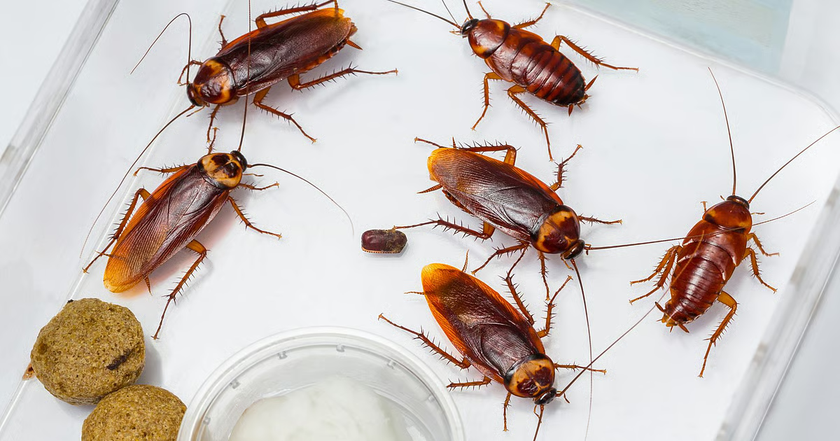




Researchers determined the atomic structure of protein crystals that form naturally inside the embryos of Diploptera punctata, the only known viviparous cockroach species. These crystals, derived from a milk-like secretion produced by the mother, pack proteins, lipids, and sugars into a highly concentrated form. A single crystal holds an estimated energy content of roughly 232 kcal per 100 g — more than three times that of an equivalent mass of dairy milk (such as buffalo milk, previously one of the densest mammalian milks at around 110 kcal/100 g) or human breast milk.
This remarkable density comes from the crystals’ composition: approximately 45% protein, 25% carbohydrates, 16–22% lipids, and additional free amino acids, making it a near-complete nutrient package.
Evolutionary Context of Nutrient Provision in Cockroaches
Diploptera punctata, commonly known as the Pacific beetle cockroach, stands out among cockroaches for its reproductive strategy. Most cockroach species are oviparous, laying eggs in protective oothecae with limited additional support. In contrast, this species is viviparous: it nourishes live embryos directly inside a brood sac for an extended gestation period of about 60–70 days (compared to 2–4 weeks for typical egg-laying species).
The mother secretes a protein-rich, yellowish fluid through the brood sac lining. Embryos ingest this fluid, which supports rapid development and leads to a substantial (over 60-fold) increase in body protein during the embryonic stage. The ingested fluid crystallizes in the embryo’s midgut, providing a stable, concentrated storage mechanism that releases nutrients gradually as needed. This adaptation allows for fewer but larger, more developed offspring.
Earlier Observations of In Vivo Protein Crystals
Scientists had previously identified milk proteins in D. punctata as early as the 1970s, noting their lipocalin-like characteristics (a family of proteins often involved in lipid binding and transport). A 1977 study detailed the milk’s composition, and genetic analyses later revealed multiple similar peptides encoded by milk genes. Observations documented crystal formation in embryo guts, but high-resolution atomic structures from functional, in vivo-grown crystals were rare. The 2016 work built on decades of prior research by resolving these crystals at atomic detail.
Isolation and Structural Analysis of the Crystals
The research team, led by scientists including Sanchari Banerjee and Subramanian Ramaswamy at the Institute for Stem Cell Biology and Regenerative Medicine in Bangalore, collected crystals directly from the midguts of embryos. Using advanced X-ray diffraction techniques — including sulfur single-wavelength anomalous dispersion (S-SAD) phasing with lower-energy X-rays — they solved the structure at an impressive 1.2 Å resolution. This allowed visualization of individual atoms, protein folds, glycosylation sites, bound lipids, and the crystal lattice packing.
The proteins, named Lili-Mip (Lipocalin-like Milk Protein), adopt a classic lipocalin fold: a beta-barrel structure with a C-terminal alpha-helix that loosely coordinates lipids in a hydrophobic pocket (capable of holding fatty acids up to 18 carbons long, such as oleic or palmitoleic acid). Crystals form in space group P1. The team compared native in vivo crystals to recrystallized versions, noting the exceptional diffraction quality despite natural heterogeneity.
Detailed Composition and Packing of the Milk Crystals
The crystals contain a heterogeneous mixture of milk proteins with variations in amino acid sequences, N-glycosylation patterns (sugars like mannose), and bound fatty acids. These glycosylated proteins bind lipids and organize into a tightly packed lattice. Surprisingly, this heterogeneity — which would typically hinder high-quality crystallization in lab settings — does not impede formation; the crystals diffract exceptionally well.
The nutrient profile includes all nine essential amino acids, making it a complete protein source. It also contains phospholipids, cholesterol, free sugars, and other lipids. This combination creates a “complete food” that releases components steadily during digestion, providing sustained energy and building blocks for embryonic growth.
Implications Drawn by the Research Team
The authors highlight that these crystals represent a unique natural storage form. “The crystals are like a complete food — they have proteins, fats and sugars. If you look into the protein sequences, they have all the essential amino acids,” said Sanchari Banerjee, then a postdoctoral researcher and lead author.
This arrangement ensures embryos have steady access to nutrients. The study also provides a rare atomic-resolution view of protein heterogeneity within a functional in vivo-grown crystal. Researchers have since explored recombinant expression of Lili-Mip in yeast and bacteria for potential applications in nutritional supplements, fortified foods, or drug carriers, though scalability remains challenging.
Study Constraints and Scope
The structural analysis focused on crystals from a specific cockroach species, using data from limited biological samples optimized for high-resolution crystallography. The work prioritizes basic structural biology, compositional insights, and reproductive biology over large-scale nutritional testing or human applications. It does not address long-term safety, allergenicity, scalability for commercial production (e.g., harvesting from thousands of insects for small volumes), or direct comparability for human diets beyond the energy density calculation.
While popular media has hyped it as a potential “superfood,” experts emphasize it is not currently suitable or available for human consumption. Production challenges and the need for safety trials limit practical use. The findings advance understanding of insect reproduction and crystallography but do not support dietary recommendations.
Reference:
Banerjee, S., Coussens, N.P., Gallat, F.-X., Sathyanarayanan, N., Srikanth, J., Yagi, K.J., Gray, J.S.S., Tobe, S.S., Stay, B., Chavas, L.M.G. & Ramaswamy, S. (2016). Structure of a heterogeneous, glycosylated, lipid-bound, in vivo-grown protein crystal at atomic resolution from the viviparous cockroach Diploptera punctata. IUCrJ, 3(4), 282–293. DOI: 10.1107/S2052252516008903.
(This article expands on the core contributions of the 2016 structural biology paper with verified details from the study and related analyses, while remaining grounded in the original scientific claims. It offers a fascinating glimpse into an unusual natural protein crystal system in insect biology.)


