Protein folding is essential to understanding how proteins achieve their functional structures. The core idea, known as the central dogma of protein science, is that the amino acid sequence dictates the three-dimensional shape of a protein. While some proteins spontaneously fold into their native structures in solution, others struggle to refold once unfolded and may misfold or aggregate. Such proteins often require chaperones to guide them into their correct form. This raises key questions about how proteins begin to fold as they are synthesized on the ribosome, and how cotranslational folding (folding during synthesis) differs from post-translational folding (folding after synthesis).
Proteins start forming secondary structures while inside the peptide exit tunnel of the ribosome, a narrow passage that also shapes the physical and chemical environment for early protein folding. One major feature of cotranslational folding is its vectorial nature: folding starts at the N-terminus, even before the rest of the protein is fully synthesized. In contrast, post-translational folding occurs after the full-length protein is available, involving more complex folding pathways.
The ribosome plays a crucial role in this process by shaping the folding environment and influencing the pathway that nascent proteins follow during their synthesis. As the emerging protein moves through the ribosome’s exit tunnel, various molecular forces and constraints affect how it folds into its final structure.
Ribosome Structure and Exit Tunnel Properties
The ribosome is a complex molecular machine responsible for protein synthesis. Structurally, it consists of two subunits: a larger 50S (in prokaryotes) or 60S (in eukaryotes) subunit, and a smaller 30S or 40S subunit. These work together to facilitate the translation of messenger RNA (mRNA) into a growing polypeptide chain.
Central to the process of cotranslational folding is the peptide exit tunnel, located within the large ribosomal subunit. This tunnel is approximately 80-100 Å long and 10-20 Å wide, with its diameter varying along its length. The narrowness of the tunnel restricts the formation of large tertiary structures but allows secondary structures, such as α-helices, to form. The chemical environment inside the tunnel is primarily hydrophilic, offering limited opportunities for hydrophobic collapse, which is a key driving force in protein folding.
The exit tunnel is not a passive conduit; its structure and internal features—such as hydrophobic patches, electrostatic charges, and ribosomal proteins—can interact with the nascent chain, subtly influencing how the protein begins to fold. For example, certain residues on the ribosome’s tunnel wall can temporarily interact with emerging peptide sequences, affecting the folding trajectory of the nascent protein.
Moreover, ribosome-associated factors like molecular chaperones often bind near the tunnel exit. These factors assist in early folding events and prevent the aggregation or misfolding of nascent chains. As a result, cotranslational folding is tightly regulated and dependent on both the physical structure of the ribosome and the activity of associated folding factors.
Stages of Cotranslational Folding
Cotranslational folding occurs in stages as the polypeptide chain is synthesized and emerges from the ribosomal exit tunnel. The process begins with the nucleation of secondary structures and progresses to the formation of more complex tertiary structures as more of the protein is synthesized.
1. Early Secondary Structure Formation
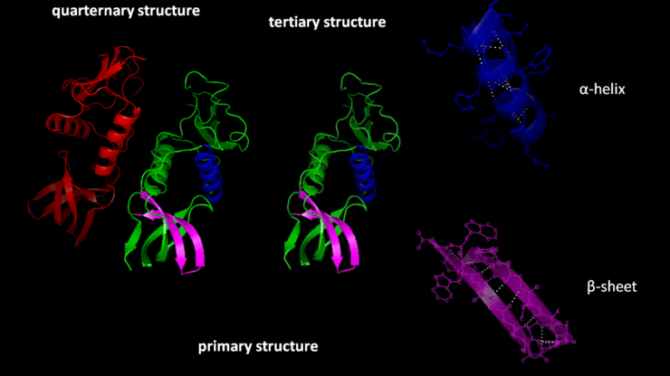
When a segment of the nascent chain emerges from the ribosome, it often forms elements of secondary structure like α-helices or β-sheets. The dimensions of the ribosomal exit tunnel allow for α-helix formation, but more complex arrangements are hindered until the chain has more room to expand.
2. Formation of Local Tertiary Structures
As the nascent chain continues to elongate, regions that have already emerged from the exit tunnel can start folding into tertiary structures, driven by interactions such as hydrophobic clustering, hydrogen bonding, and electrostatic forces. This stage often involves the folding of domains, which are independent, functional regions within proteins. Many proteins fold in a domain-by-domain manner as their synthesis proceeds.
3. Assistance by Molecular Chaperones
Once the nascent chain reaches the cytoplasm outside the ribosome, molecular chaperones like Trigger Factor (TF) in prokaryotes or NAC (Nascent polypeptide-Associated Complex) in eukaryotes assist in preventing misfolding or aggregation of the emerging chain. These chaperones stabilize unfolded or partially folded proteins, giving them time to reach their correct conformation.
4. Completion of Folding Post-Translation
While much of the folding process can be completed during translation, some proteins, especially larger and more complex ones, may not reach their fully folded state until after synthesis is complete. These proteins may require the intervention of additional chaperones, such as Hsp70 and Hsp60, which assist in the folding of newly synthesized proteins or refold misfolded ones.
Influence of the Ribosome on Protein Folding
The ribosome plays a significant role in guiding the folding process. The ribosomal exit tunnel provides a restricted environment, where early folding events, such as α-helix formation, are possible but limited. The ribosome also interacts with the nascent chain in ways that influence its folding path.
1. Steric Constraints in the Exit Tunnel
The exit tunnel confines the nascent chain, limiting its ability to fold into large structures while still inside. Small motifs like α-helices can form, but complex tertiary structures typically cannot be accommodated until the chain has emerged further. This gradual exposure of the nascent chain allows proteins to fold in a sequential manner, which can prevent misfolding.
2. Interplay with the Ribosome-Associated Chaperones
Ribosome-associated chaperones, such as Trigger Factor (TF) in bacteria and NAC (Nascent polypeptide-Associated Complex) in eukaryotes, bind to the nascent chain as it exits the ribosome. These chaperones play a key role in shielding exposed hydrophobic regions and preventing aggregation. They also provide a stabilizing influence that promotes correct folding as the nascent chain elongates.
3. Ribosome as a Platform for Efficient Folding
The ribosome’s structure and interactions with the nascent chain suggest that it provides more than just a passive environment for protein synthesis. It serves as a platform for efficient folding by spatially organizing the emerging chain and allowing it to fold incrementally. Some studies suggest that the ribosome can even catalyze specific folding events by promoting certain structural formations.
Role of Molecular Chaperones in Protein Folding
Molecular chaperones are essential to ensure that newly synthesized proteins achieve their proper three-dimensional structure, avoiding misfolding and aggregation. They bind to nascent polypeptide chains during translation or to proteins that are partially unfolded due to stress. There are several types of chaperones, each with specific roles in the folding process.
1. Hsp70 Chaperones
The Hsp70 family of chaperones is one of the most well-studied groups involved in protein folding. Hsp70s are ATP-dependent and have two major domains: a nucleotide-binding domain (NBD) and a substrate-binding domain (SBD). Here’s how they work:
- Recognition and Binding: Hsp70 binds to hydrophobic regions of the nascent chain, which are often prone to aggregation.
- ATP Hydrolysis: The binding of ATP to the Hsp70 allows the chaperone to adopt an open conformation. Once ATP is hydrolyzed, Hsp70 clamps down on the protein substrate, preventing aggregation and giving the protein more time to fold properly.
- Release and Refolding: The cycle of binding and release helps the protein reach its native conformation. Co-chaperones such as Hsp40 facilitate this process by stimulating the ATPase activity of Hsp70, while nucleotide exchange factors (NEFs) assist in the release of ADP, resetting the Hsp70 to bind another misfolded protein.
2. Hsp60/GroEL Chaperonins
Chaperonins are large, barrel-like complexes that provide a protected environment for protein folding. In bacteria, this system is represented by GroEL-GroES, while eukaryotes have the Hsp60-Hsp10 system.
- GroEL: The GroEL complex has two stacked rings, each forming a chamber. Misfolded proteins are captured in one of these chambers.
- GroES: The co-chaperone GroES acts as a lid, sealing the protein inside the chamber, where it is isolated from the cellular environment.
- ATP-Driven Folding Cycle: Inside the chamber, ATP binding and hydrolysis induce conformational changes that give the protein the opportunity to fold correctly. This process may involve multiple rounds of encapsulation and release, increasing the chances of the protein adopting its correct structure.
3. Hsp90 Chaperones
The Hsp90 family plays a specialized role in the folding of regulatory proteins such as kinases and steroid hormone receptors. It works in tandem with co-chaperones and is also ATP-dependent.
- Client Proteins: Hsp90 primarily works with more complex and larger proteins that require precise folding for their function.
- Folding Process: Hsp90 binds to client proteins in their near-native state, facilitating the final stages of folding. This process is tightly regulated, and ATP hydrolysis drives the structural rearrangements necessary for folding completion.
4. Small Heat Shock Proteins (sHSPs)
Small heat shock proteins (sHSPs) function differently from the ATP-dependent chaperones. They act as holding chaperones

