The heart originates from a bilateral structure in the embryo. There, mesenchymal cells form a pair of endocardial tubes below the pharynx. These tubes fuse into a single endocardial tube, which, along with the surrounding splanchnic mesoderm, forms the heart. This two-layered tube consists of an inner endocardium and an outer myocardium. The heart becomes S-shaped due to differential growth and the formation of chambers and valves.
Single-Chambered and Primitive Hearts
Primitive Chordates: Absence of a True Heart
Amphioxus, a primitive chordate, lacks a true heart. Instead, a contractile part of the ventral aorta beneath the pharynx acts as a heart. This primitive heart is enclosed within a pericardial cavity, which a transverse septum separates from the body cavity. The conus arteriosus connects to the ventral aorta and functions during circulation.
The Two-Chambered Heart in Fish
Cyclostomes
In Cyclostomes, the heart has four chambers arranged sequentially: a thin-walled sinus venosus, a slightly muscular auricle, a muscular ventricle, and a muscular conus arteriosus and bulbus cordis. These chambers lie within the body cavity alongside other visceral organs. Evolution has brought significant changes to the heart’s structure.
Elasmobranchs (Cartilaginous Fish)
In elasmobranchs (cartilaginous fish like dogfish), the heart is a muscular, dorsoventrally bent S-shaped tube with four compartments in a linear series. The sinus venosus and auricle receive venous blood, and the ventricle and conus arteriosus pump blood. We call this heart a branchial venous heart because only deoxygenated blood is pumped to the gills, where it gets oxygenated and then distributed to the body.
Teleosts (Bony Fish)
The heart in teleosts resembles that of elasmobranchs but with some differences. The conus arteriosus is reduced and contains a single pair of valves. The ventral aorta’s proximal part, the bulbus arteriosus, is thick-walled and elastic, which aids in blood circulation during ventricular contraction.
Three-Chambered Hearts: Transition to Double Circulation
Lungfish
In lungfish, a septum divides the atrium into right and left chambers, reflecting a shift towards a double circulatory system where both oxygenated and deoxygenated blood enter the heart separately. The right auricle receives deoxygenated blood and pumps it to the gills or lungs, while the left auricle receives oxygenated blood returning from the lungs.
Amphibians
Amphibians show a further evolution of the heart, where the atrium is completely divided into right and left chambers. The conus arteriosus divides into systemic and pulmonary vessels by a spiral valve, which facilitates better separation of oxygenated and deoxygenated blood. However, some mixing still occurs, especially in lungless salamanders where the interatrial septum is incomplete.
Reptiles: Ventricular Partitioning
In reptiles, the heart has advanced further with a complete separation of the atrium into right and left chambers. The ventricle is partially divided, with complete separation in alligators and crocodiles. This structure ensures better separation of oxygenated and deoxygenated blood. The embryonic conus arteriosus splits into the pulmonary arch and systemic aorta, enhancing the efficiency of blood circulation.
Four-Chambered Heart in Birds and Mammals: Complete Separation
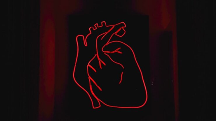
In birds and mammals, the heart is fully divided into four chambers: two atria and two ventricles. This complete separation allows for efficient double circulation without any mixing of oxygenated and deoxygenated blood. The systemic aorta leaves the left ventricle, supplying oxygenated blood to the body, while the pulmonary artery leaves the right ventricle, carrying deoxygenated blood to the lungs for oxygenation. The sinus venosus is incorporated into the right atrium, enhancing the efficiency of venous return. This design represents the peak efficiency in the vertebrate heart’s evolution, supporting the high metabolic demands of endothermy.


