Alimentary Canal in Vertebrates
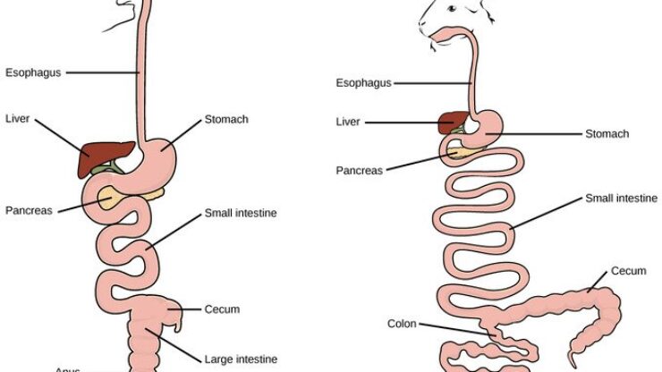
The alimentary canal, also known as the digestive tract or gastrointestinal (GI) tract, is a long, tube-like structure that extends from the mouth to the anus.The alimentary canal is the pathway (a tube-like structure) that starts from the mouth and ends in the anus. It plays a primary role in the digestion of food. The canal is divided into several regions, including the mouth, esophagus, stomach, small intestine, and large intestine
STUDY ZONE
The alimentary canal, also known as the digestive tract or gastrointestinal (GI) tract, is a long, tube-like structure that extends from the mouth to the anus. The alimentary canal is the pathway (a tube-like structure) that starts from the mouth and ends in the anus. It plays a primary role in the digestion of food. The canal is divided into several regions, including the mouth, esophagus, stomach, small intestine, and large intestine, each specialized for specific functions such as mechanical breakdown, enzymatic digestion, nutrient absorption, and waste elimination.
Alimentary Canal in Vertebrates
In vertebrates, the alimentary canal is a continuous tube starting from the mouth and ending at the anus. It is responsible for the digestion and absorption of food. The process of digestion often begins in the buccal cavity and involves further breakdown of the bolus, absorption of nutrients, and elimination of waste.
Alimentary Canal Design
The design of the alimentary canal reflects the diet of the organism and varies among phylogenetically related vertebrates. Despite these differences, the canal typically consists of the esophagus, stomach, intestines, and cloaca, all sharing a common structural design.
Common Plan of the Alimentary Canal
The alimentary canal is a hollow tube with four main layers:
1. Mucosa: The innermost layer, containing epithelium, muscularis mucosae, and lamina propria.
2. Submucosa: A layer of loose connective tissue with nerve plexuses.
3. Muscularis Externa: Consists of circular and longitudinal smooth muscle layers.
4. Adventitia: Fibrous connective tissue, which becomes the serosa when enveloped by the mesentery.
Embryonic Development
Endoderm: Forms the lining of the gut.
Mesoderm: Forms the smooth muscles, connective tissues, and blood vessels.
The development involves twisting and coiling to fit the gut into the body cavity without kinking.
Gut Development Stages
1. Formation of the primary gut loop.
2. Elongation and twisting in the first major coil.
3. Further elongation and coiling to create a compact digestive tube.
4. Differentiation into distinct digestive regions.
Stomach
Structure and Function
1. Stomach Function and Structure:
- Receives bolus from the esophagus.
- Expanded region absent in cyclostomes and protochordates.
2. Stomach in Urochordates:
- Present in some urochordates.
- Receives mucus with food particles from the branchial basket.
3. Storage Component:
- Stores large quantities of food for carnivores.
- Allows time for mechanical and chemical digestion to commence.
4. Role of Hydrochloric Acid:
- Produced by the stomach to retard food putrefaction.
- Preserves food until digestion begins.
5. Digestion Processes:
- Absorption of water, salts, and vitamins.
- Churns and mixes food with gastric juice.
6. Gastric Juice Composition:
- Contains enzymes and mucus.
- Primarily composed of hydrochloric acid.
7. Stomach Morphology:
- Expanded size differentiates it from the esophagus and small intestine.
- Internal wall forms rugae when not distended.
8. Histological Character of the Mucosal Wall:
- Used to distinguish functional regions within the stomach.
Functionality During Digestion
- The gut becomes mechanically and chemically active with churning, peristaltic, and digestive secretions.
- Activity controlled by the mesenteries.
Esophagus
Function
1. Function of Esophagus:
- Connects the pharynx to the stomach.
- Acts as a transport tube for food entering the mouth through the throat into the stomach.
2. Mucous Secretion:
- Assists smooth passage of food.
- Yearly produces enzymes for chemical digestion.
3. Esophageal Mucosa:
- Lined with ciliated cells in some vertebrates to control mucous flow.
- Helps in mixing small crumbs in the stomach.
4. Types of Epithelium:
- Keratinized epithelium in some vertebrates for rough food.
- Ciliated epithelium in others for mucus.
5. Temporary Storage:
- Serves as a storage site for large quantities of food before digestion.
6. Muscle Composition:
- Straight muscle anteriorly transitioning to smooth muscle posteriorly.
Intestine
Features
- Intestinal Mucosa Features:
- Contains an epithelium with numerous microvilli.
- Microvilli increase absorption surface area and create a microenvironment for digestive enzymes.
- Digestive enzymes bound to microvilli include disaccharidases, peptidases, and lipases.
Intestinal Glands (Crypts of LieberkΓΌhn)
- Serve as a reservoir for immature cells that mature into absorptive epithelial cells.
Small Intestine
- Composed of the duodenum, jejunum, and ileum.
- Duodenum receives chyme and exocrine secretions from the liver and pancreas.
- Jejunum and ileum have distinct histological features.
Large Intestine
- Larger in diameter, lacks villi, and may form a loop called the colon.
- Ends in a rectum or cloaca, depending on whether it recovers only alimentary or also urinary/reproductive products.
Functions
- Peristaltic movements push food along.
- Adds secretions for digestion and lubrication.
- Absorbs final products of digestion and water.
Accessory Glands
- Duodenal Glands: Neutralize acidic stomach chyme.
- Pancreas: Releases proteolytic enzymes into the duodenum.
Cloaca
- The cloaca lies at the end of the embryonic gut from the proctodeum.
- Acts as a common chamber for products from the intestine and urogenital tracts.
- Present in some fishes, but most mammals do not have a cloaca.
Specializations of the Alimentary Canal
Length and Structure
1. Longest Digestive Path:
- Spiral valve increases the root length in the alimentary canal.
- Partition prolongs digestion by making food wind through a spiral channel.
- Carnivores have a short intestine, and herbivores have a long intestine for nutrient extraction.
2. Larval Lampreys:
- Typhlosole, a longitudinal fold, increases the surface area for absorption.
- Site of hematopoiesis (blood formation) in larvae.
- In adults, replaced by numerous folds to increase absorptive area.
3. Expansions and Extensions:
- Crop: A baglike esophageal expansion in birds for food storage.
- Cecum: A blind-ended outpouching at the junction of the small and large intestine.
4. Differentiation and Regionalization:
- New regions may form, differentiating into the small and large intestines.
- The cloaca is lost as separate outlets develop for the intestine and urogenital tracts.
Vascularization of the Gastrointestinal Tract
Blood Supply
1. Origin:
- Supplied to the gut via the dorsal aorta, with major branches like the celiac and mesenteric arteries.
2. Sequential Supply:
- Supplies sections supporting active segments of the gut while reducing flow to inactive segments through peripheral vasoconstriction.
3. Arterial Distribution:
- Arteries penetrate the gut wall to reach the submucosa.
- In the submucosa, they form a plexus and distribute to the muscular layer and mucosa.
4. Capillary Beds:
- Extensive capillary beds in the mucosa support active absorption in the small intestine.
- Facilitate the transport of digested nutrients out of the gut.
5. Lymphatic System:
- Accompanies blood vessels, forming an extensive network of lymphatic capillaries in the mucosa.
- Lymphatic vessels, especially lacteals in the small intestine, are crucial for the absorption of long-chain fatty acids.


Digestive Systems of Various Fish and Animal Species
Ostracoderm Fishes
Digestive System: Little is known about their digestive systems, inferred from coprolites (fossilized feces).
Diet: Detritus from the substrate.
Intestine: Small intestine suggested by the coiled structure of coprolites.
Cyclostomes
Alimentary Canal: A straight tube from mouth to anus without coelom or major bends.
Esophagus: Ciliated, runs directly from the pharynx to the intestine. The stomach is absent.
Diet: Includes small particulate matter, blood, tissue, and detritus.
Food Passage: Direct from esophagus into the intestine.
Lampreys
Metamorphosis: A new esophagus forms during metamorphosis.
Pharynx: Used for tidal ventilation.
Intestine:
- Anterior Section: Absorption of fat, osmoregulation in marine environments.
- Posterior Section: Protein absorption, elimination of biliverdin (a bile pigment).
Liver Products: Enter the anterior end of the intestine via bile ducts.
Gnathostome Fishes (Jawed Fishes)
Variations in Alimentary Canal Design:
- Common Features: Esophagus, stomach, and intestine.
- Stomach: Usually J-shaped, with a wide fundic region and a narrow pyloric region.
- Spiral Valve: Found in the intestines of elasmobranchs and some bony fishes, absent in teleosts (modern bony fishes).
Teleosts (Modern Bony Fishes):
- Intestine: Elongated, often folded back on itself.
- Terminal Section: Usually widened into the cloaca or rectum.
Pyloric Caeca:
- Open into the duodenum.
- Primary areas for digestion and absorption.
Tetrapods
Amphibians:
- Esophagus:
Short, transitions gradually to the stomach.
- Stomach Mucosa:
Features gastric glands, including fundic glands and pyloric glands near the passage to the intestine.
- Intestine:
Differentiates into a coiled small intestine (beginning with the duodenum) and a short, straight large intestine ending in the cloaca.
Reptiles:
- Alimentary Canal:
Similar to amphibians but larger and more elaborated in some species.
- Stomach:
Many lizards have a heavy-walled, muscular stomach. Crocodiles and alligators have a gizzard with thick muscles for grinding food against ingested stones.
- Gastric Juice:
Produced in the glandular region of the crocodilian stomach.
- Large Intestine:
Typically distinct, with some herbivorous lizards having a cecum between the small and large intestines.
Birds:
- Esophagus:
Forms an inflated crop for temporary food storage. In species like pigeons, it secretes a nutritional fluid known as "milk" for the young.
- Stomach:
Consists of a thin-walled glandular section (proventriculus) and a posterior gizzard, which aids in grinding down food.
- Intestine:
Long, with a small intestine that includes the duodenum and ileum, and a short large intestine that empties into the cloaca. One or several caeca often sprout near the junction of the large and small intestines.
Mammals:
- Stomach and Esophagus:
Expanded into a pouch for temporary food storage.
- Small Intestine:
Long and coiled, distinguishable into the duodenum, jejunum, and ileum.
- Large Intestine:
Often long but not as small as the small intestine.
- Herbivorous Mammals:
Usually have a cecum at the junction of the small and large intestines. In humans, this is referred to as the vermiform appendix.
Drop Us a Line
Weβd Love to Hear from You