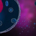
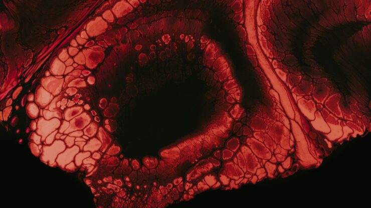
The Fascination with Oocytes because of their remarkable ability to remain viable for up to decades, oocytes—the germ cells that give rise to eggs—have long been of great interest to researchers. Although a cell arrives prenatally, it continues to function even though virtually all of its cellular components are replaced multiple times per year. This has begged explanation regarding how these cells persist with relatively minimal damage over time.
Insights from New Research
A new study, led by biochemist Melina Schuh at the Max Planck Institute for Multidisciplinary Science, has shed new light on the mystery: mammalian ovaries harbor proteins with lives that are astonishingly long. Results published in Nature Cell Biology offer more detailed insight into how oocytes maintain their integrity throughout an animal’s reproductive life and what contributes to fertility decline when the ovaries age.
Long-Lived Proteins in Oocytes
It has previously been known that in the aging process there are long-lived proteins; however, this paper is the first to isolate specifically to characterize these proteins in the ovary. Although the biology of extremely long-lived proteins in aging has been known for a while, this is the first paper to carefully characterize the nature and identity of those proteins in the ovary, says Lei Lei, a reproductive biologist at the University of Missouri School of Medicine, who was not involved in the study. It is relevant in sustaining new life because oocytes cannot afford mistakes in their molecular makeup to preserve the structure and function of these proteins perfectly.
To figure out the lifespan of proteins in oocytes, Schuh’s group thought of a pretty clever scheme. They fed the pregnant mice a diet that contained an amino acid rich in a heavy isotope of carbon, which was incorporated into the proteins of both the mothers and their in utero pups. Then at the time of delivery, they switched the pups over to a diet that contained a lighter isotope of the same amino acid, allowing them to follow which proteins were synthesized prenatally and for how long they persisted. At puberty, the researchers quantified these mouse oocytes’ proteins by mass spectrometry and found that about 10% of them were produced before birth. Such long-lived proteins comprised those involved in essential cellular structures like mitochondria, ribosomes, and chromatin and played a critical role in metabolism and DNA repair.
Ovarian Somatic Cells and Protein Longevity
While not oocytes, the somatic cells of the ovary do harbor long-lived proteins. Beyond the dendritic cells, the study also examined other types of ovarian cells—including stromal and thecal cells—important for female fertility. Even in these somatic cells, the authors noted that more than 10% of the proteins had half-lives exceeding 100 days, with many proteins lasting throughout most of the animals’ lives. Interestingly, there were many more long-lived proteins in the ovary than in other tissues, including cartilage, brain, and muscle, where only <1% of proteins survived for such extended periods.
Preservation of Oocytes from Protein Misfolding
One of the big questions that Schuh’s team had was how these proteins survive degradation for so long. One place where perhaps there was a clue was with the examination of protein homeostasis, or the function by which cells regulate protein synthesis and degradation. Notably, no evidence for protein misfolding and aggregation was observed in aged oocytes, and the activity of proteasomes—that is, protein complexes that degrade damaged proteins—remained high even in older oocytes. This would signify that these cells have robust mechanisms that negate the likelihood of protein misfolding and protect against oxidative damage, thus ensuring long-term presence of these crucial proteins.
Moreover, the ovary had a high level of antioxidants and protein chaperones—molecules that assist in protein folding. This makes the existence of highly potent systems that help oocytes maintain the stability of proteins for a long period more likely.
Decline with Aging and Fertility
Even with these protective mechanisms in place, ovarian aging necessarily impacts protein composition. Analyzing mice of all ages, from one day to 11.5 months, Schuh’s group reported a tremendous, age-related depletion of long-lived proteins. Such depletion could be driving widespread remodeling of ovarian proteins, perhaps part of the reason fertility often declines so precipitously after three months of age in mice.
Implications for Human Fertility
It’s a mouse study, but the implications for human ovarian biology are interesting. “The same long-lived proteins should exist in human ovaries,” says Schuh. And not long from now, researchers will indeed look into this phenomenon in people. “How these results relate directly to humans, we do not know yet,” Schuh says, but she hopes that future research may expand our knowledge of the processes going on in humans, potentially bringing new insights into fertility and ovarian aging.
The discovery of long-lived proteins within the ovary now opens an entirely new route for exploring this possibility of preserving reproductive cells for a longer duration. However, Lei points out that it would be unwise to extract direct medical therapy from these discoveries: “Human ovarian biology is much more complex than in mice. However, an understanding of these proteins and their role in fertility might someday provide clues for how to address age-related fertility decline in women.”
References
- Harasimov K, et al. Extensive protein longevity underlying maintenance of oocytes in mammalian ovary. Nat Cell Biol. 2024;26(7):1124–1138.
- Tachibana-Konwalski K, et al. Rec8-containing cohesin complexes are required for bivalent chromosome maintenance during the prophase I progression of mouse oocytes. Genes Dev. 2010;24(22):2505–2516.