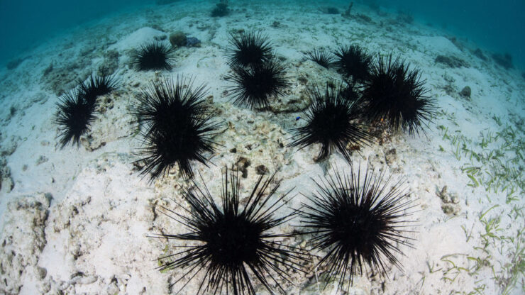
Sea urchin twinning has fascinated scientists for more than a century, raising the question of how two complete individuals can emerge from a single fertilised egg. For over a century, scientists have been puzzled by a remarkable biological phenomenon: how can two complete individuals emerge from a single fertilised egg?
This new research from the University of Tsukuba, published in Nature Communications, has shed light on this long-standing question, revealing the intricate cellular and molecular mechanisms behind sea urchin twinning.
The study, drawing on Hans Driesch’s pioneering experiments from the late 19th century, details how early-stage sea urchin embryos can self-organise and regenerate their entire developmental plan even after being split in half.
Unique Developmental Trajectory
The researchers focused on the Japanese sea urchin species, Hemicentrotus pulcherrimus, and found that when a 2-cell stage embryo is bisected, each isolated half undergoes a unique and previously unreported developmental process. Unlike intact embryos that directly form a blastula, these halved embryos first develop into a flat, plate-like structure, then transition into a cup-like shape, and finally close to form a sphere. This spherical structure eventually develops into a miniature, yet complete, blastula. This distinct sequence of “flat, cup, and sphere” stages was also observed in S. purpuratus and Strongylocentrotus intermedius, though interestingly, Temnopleurus reevesii embryos develop directly into a blastula without these intermediate shapes. This suggests potential species-specific variations in regulative development.
The Mechanics of Shape Transformation
The dramatic shift from a flat to a spherical shape is not driven by increased cell proliferation, but rather by dynamic cell shape changes. The study identified two key molecular mechanisms facilitating this morphological reorganisation. Firstly, actomyosin activity plays a crucial role. Live imaging revealed strong actin polymerisation on the basal side of cells during the cup-to-sphere transition, leading to cell elongation along the apicobasal axis, giving cells a distinct cone-like shape. Inhibiting actin polymerisation with cytochalasin D or myosin-II ATPase with (-)-blebbistatin prevented or delayed proper sphere formation, highlighting the essential role of actomyosin-generated forces.
Secondly, septate junctions, which act as occluding junctions in invertebrates, contribute significantly to this shape transition. The timing of the flat-to-cup transition coincided with the initiation of septate junction formation. Knockdown experiments targeting tetraspanin and ZO-1, genes related to septate junctions, resulted in “bumpy” flat shapes where cells failed to form a single layer and struggled to achieve a spherical form. These findings suggest that the adhesive forces provided by septate junctions enable the edges of the cup-like structure to bind and close, forming a cohesive sphere. Crucially, the researchers concluded that these morphological changes are largely driven by cell-autonomous mechanisms, not by major signaling pathways typically associated with body axis formation.
How the Body Finds Its Axes Again
Beyond forming a proper spherical shape, the halved embryos face the challenge of re-establishing their body axes. The study revealed a temporary disorganisation of the anterior-posterior (A-P) axis immediately after sphere formation, with anterior (foxQ2) and posterior (foxA) marker genes expressed in close proximity. However, this disruption is transient; within hours, the A-P axis is reorganised to resemble normal patterning. This re-organisation primarily involves the shifting of the anterior end specification site from its original position, while the posterior end largely maintains its location.
The Wnt/β-catenin signaling pathway was found to be essential for this anterior shift. Live imaging showed temporal nuclear β-catenin signals adjacent to the original posterior site after sphere formation, and inhibition of Wnt signaling prevented the anterior shift. This suggests that Wnt/β-catenin, while typically not restricting the anterior end in intact embryos at this stage, is reactivated to facilitate axis re-formation in halved embryos. Non-canonical Wnt signaling also partially contributes to this process.
The dorsal-ventral (D-V) axis also undergoes re-organisation. Initially, key D-V patterning genes like nodal are broadly expressed across the embryo during the cup and sphere stages, unlike their typical biased expression. However, after sphere formation, nodal expression re-biases towards the future ventral side, restoring normal D-V patterning. The re-establishment of the A-P axis appears to indirectly trigger D-V axis formation, possibly by clearing the Nodal repressor FoxQ2, thereby allowing the stable activation of the Nodal autoregulatory loop necessary for robust D-V axis formation.
Broader Impact on Life Sciences
This research not only provides the first experimental evidence of the molecular mechanisms underlying axis re-formation during self-organization but also highlights the robustness and flexibility of embryonic developmental programs. The ability of sea urchin embryos to autonomously re-establish their developmental axes and molecular gradients after disruption offers crucial insights into regulative development and the origins of monozygotic twinning in humans. The parallels observed with Xenopus laevis suggest conserved evolutionary pathways for self-organisation. These findings could have significant implications for regenerative biology and artificial tissue engineering, where harnessing such self-organising principles might be key for tissue repair and organ regeneration.
REFERENCE
Suzuki, H., Yaguchi, J., Tsuyuzaki, K., & Yaguchi, S. (2025). Unraveling the regulative development and molecular mechanisms of identical sea urchin twins. Nature Communications, 16, 8005.

